
Abstract
Biofluorescence in echinoderms is largely unexplored, and even though the green sea urchin Strongylocentrotus droebachiensis is a well-studied species, the presence and/or function of fluorescence remains very poorly understood. Hyperspectral imaging was conducted on adult sea urchins (N = 380) while fluorospectrometric analysis was conducted on sea urchin coelomic fluid (N = 30). Fluorescence was documented in both the spines and coelomic fluid of S. droebachiensis. Intact spines exhibited a low intensity green emission (∼550–600 nm), while broken spines averaged a high emission peak in the green spectrum (∼580 nm). Sea urchins produce a red exudate with a pronounced emission peak (∼680 nm) with a shoulder peak (∼730 nm). The sampled coelomic fluid exhibited high variability, with a majority exhibiting a low-level green fluorescence while pronounced emission peaks (N = 5) were found in the red spectrum (∼680 nm). The complex fluorescence produced by S. droebachiensis warrants further investigation on its applicability for monitoring welfare of sea urchins in aquaculture facilities.
Export citation and abstract BibTeX RIS

Original content from this work may be used under the terms of the Creative Commons Attribution 4.0 licence. Any further distribution of this work must maintain attribution to the author(s) and the title of the work, journal citation and DOI.
1. Introduction
Marine organisms produce biofluorescence through the uptake of photons from the spectrally restricted (blue-shifted) environment and emit a lower, less energetic wavelength light via fluorochromes present in their bodies [1]. Several families of invertebrates have been documented producing biofluorescence, with the two-step production of light in hydrozoan medusa Aequorea Victoria perhaps the most well-known. The chemiluminescent protein aequorin emits blue light (470 nm) in response to calcium ion bonding, which is subsequently reemitted by the Green Fluorescent Protein (GFP) as a green fluorescence (508 nm) [2–4]. Biofluorescence in echinoderms is largely unexplored and even though Strongylocentrotus droebachiensis is a well-studied species, the presence and/or function of fluorescence remains very poorly understood.
The green sea urchin S. droebachiensis is a commercially valued echinoid within the circumpolar northern hemisphere [5]. The biomass of S. droebachiensis along the Norwegian coastline is estimated to be 80 billion individual animals/56,000 tons [6]. The poor quality of roe from wild caught urchins in Norway means there is a need for development of echinoculture methods in aquaculture facilities that can enhance the roe quantity and quality and ensure consistent quality for market consumption. An important limiting factor in echinoculture is the lack of background data and associated diagnostic techniques for monitoring their health and welfare. Clinical disease in sea urchins is poorly understood [7–9]. Echinoderms such as S. droebachiensis are susceptible to acute and chronic infections by microbial pathogens and parasitic nematodes [10]. In the northwest Atlantic, periodic mass mortalities caused by Paramoeba invadens can have impact on populations [10, 11]. Disease outbreaks and mass mortalities amongst wild echinoids are increasing over the last few decades [9, 12, 13]. In addition, there is very limited literature on how stress is manifested in sea urchin physiology and how such stress could be measured accurately within echinoderms. As aquaculture operations such as capture and captive holding are known to expose organisms to both acute and chronic stress [14], biological functions such as somatic growth and maintenance [15], reproduction [16], and immune response [17] typically suffer as the organisms' physiology reallocate resources to stress adaptation. Successfully monitoring sea urchin health and welfare is vital for future aquaculture activities as sea urchins are prone to sudden mortality events in aquaculture. Our study aims to evaluate biofluorescence in S. droebachiensis within a controlled setting and whether such fluorescence can be considered a dynamic process that can be monitored through hyperspectral or fluorospectroscopy technology as a non-invasive means of measuring the physiological status of S. droebachiensis
2. Materials and methods
Green sea urchins were collected in Kvalsund, Norway, (69° 45' 16.5312' N, 19° 2' 2.0904' E) between 10–12/2022 (figure 1). Urchins were collected by free divers in net bags and placed in a seawater filled container (1,000 liters) within minutes of being removed from seawater for transport to the NOFIMA laboratory facilities (Tromsø, Norway). A total of 380 urchins were scanned on arrival at the facility. Of those animals, a random sample of 30 sea urchins were sampled for coelomic fluid for fluorospectroscopy.
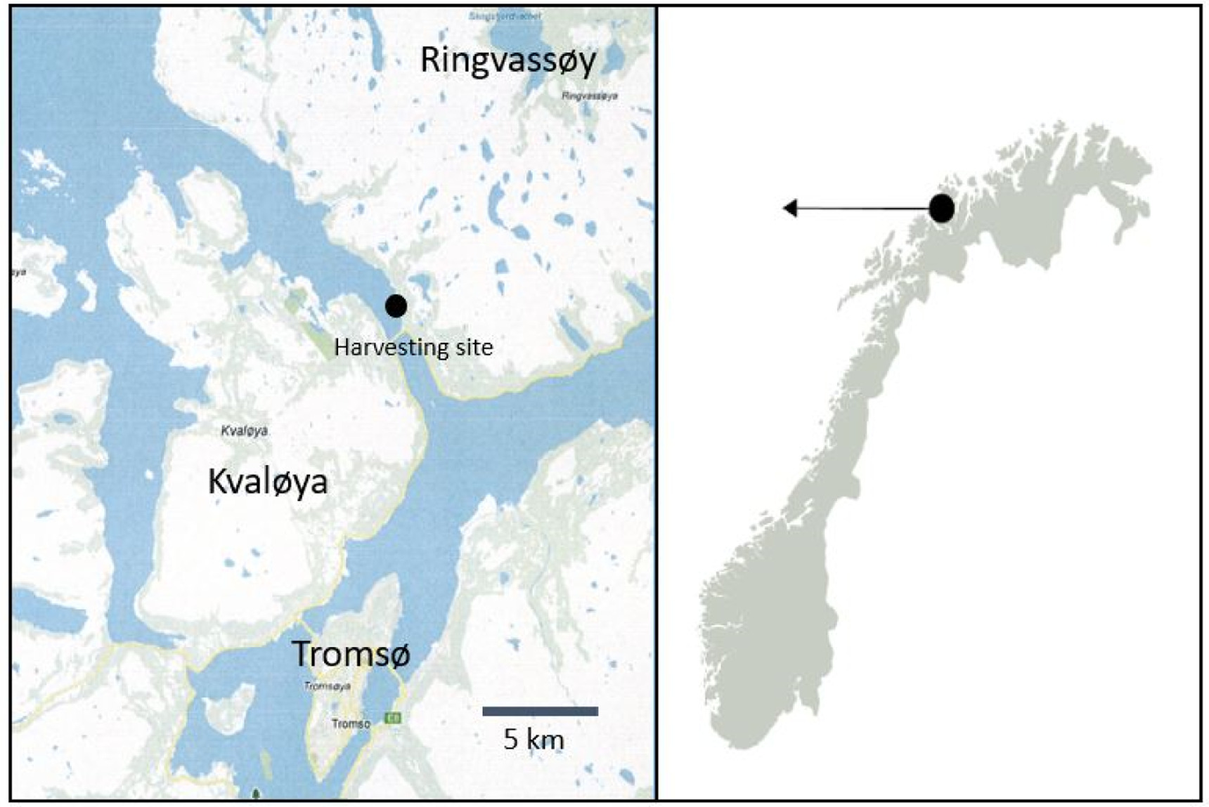
Figure 1. Green sea urchins Strongylocentrotus droebachiensis were collected from Kvalsund region of Northern Norway.
Download figure:
Standard image High-resolution imageFor sampling, the sea urchins were randomly assigned a numbered square on a Styrofoam lid for scanning on a conveyor belt set at 44 mm s−1 to yield an along track resolution equal to twice the across trac resolution. The conveyor belt was illuminated by the G5 XR30 Pro Radion LED lighting (Ecotech Marine Bethlehem, PA USA) in royal blue (∼445 nm). Hyperspectral imaging was taken with the HySpex VNIR-1800 hyperspectral camera (Norsk Elektro Optikk AS, Oslo, Norway), with a spectral range from 400 to 1000 nm, a spectral resolution of 5.5 nm, and 1800 spatial pixels. The camera was mounted 1 m above the conveyor belt, resulting in a field of view of 300 mm. The spatial resolution across the track was 0.17 mm, with 1800 pixels across the conveyor belt. The images were spatially binned in the across track direction to yield square pixels with a resolution of 0.34 mm.
Red, green, and blue (RGB) photographs for illustrative purposes were taken using a digital single lens reflex (DSLR) camera (D5100, Nikon, USA/Nikon AF-S 60 mm f/2.8 G IF-ED Micro lens) and a yellow barrier filter (Tiffen 62DY15 62 mm Deep Yellow 12 Filter). A 20-liter photographic aquarium constructed of optic white glass with a moveable black acrylic background was used to take RGB photographs of sea urchins within sea water.
Coelomic fluid (2 ml) was extracted from the peristomal membrane of sea urchins using a 26-gauge hypodermic needle attached to a sterile syringe (BD microlance, Eysins, Switzerland). Each syringe was pre-loaded with 0.5 ml of pre-chilled citrate/EDTA anticoagulant based on a formula for crustaceans [18]) [0.45 M NaCl, 0.1 M glucose, 30 mM sodium citrate, 26 mM citric acid, 10 mM EDTA; pH 5.4]. Syringes containing coelomic fluid were placed on crushed ice prior to analysis. A Duetta fluorescence and absorbance spectrometer (HORIBA Scientific, Kyoto, Japan) was used to analyze hemolymph samples placed within a 3.5 ml fluorescence quartz cuvette (Science Outlet Inc., Weifang, China). The recorded fluorescence emissions were analyzed within the EZ Spec Software (HORIBA Scientific, Kyoto, Japan). The excitation wavelengths from 250 to 350 nm were recorded and emission wavelengths from 400 to 800 nm with a 0.5 nm spectral resolution.
Raw data files collected with hyperspectral scanning were radiometrically calibrated with HySpex Rad v2.5 (Norsk Elektro Optikk, AS, Oslo, Norway) to produce radiance values in units of  The radiometrically calibrated data was uploaded for analysis in Breeze hyperspectral imaging software (Prediktera, Umeå, Sweden) and each sea urchin was cropped out into a separate image. A 33 × 33 grid was then applied, and each square in the grid was treated as a sampling point to distinguish different spectral features in the sea urchins in a Principal Component Analysis (PCA) plot.
3. Results
Fluorescence was documented in both the spines and coelomic fluid of S. droebachiensis (figure 2). Intact spines exhibited a low intensity green emission (∼550–600 nm), while broken spines averaged a high emission peak in the green spectrum (∼580 nm). Green sea urchins also produce a red exudate with a pronounced emission peak (∼680 nm) with a shoulder peak (∼730 nm). A PCA plot of the standard normal variate (SNV) transformed mean spectra (figure 3) shows clusters of samples based on their similarity; strong clustering was detected within the strong green fluorescence from broken spines, the weakly green fluorescence from intact spines, and the strong red fluorescence from exudate.

Figure 2. Results of external hyperspectral analysis of green sea urchins (N = 380). Intact spines exhibited a broad low green emission (∼550–600 nm) (a), while broken spines averaged a dominant peak in the green spectrum (∼580 nm) (b). A pronounced peak (∼680 nm) (c) with a shoulder peak (∼730 nm) (d) was documented in a red exudate produced by urchins.
Download figure:
Standard image High-resolution image
Figure 3. PCA plot of SNV-transformed mean spectra from a 33 × 33 grid over each sea urchin image. 1: strong green fluorescence from broken spines, 2: weakly green fluorescence from intact spines, 3–5: background pixels. 6: strong red fluorescence from exudate. The t(2) and t(3) are the second and third principal components of the spectra. The color bar represents the point density, where each point is a square in the grid placed over images.
Download figure:
Standard image High-resolution imageThe sampled coelomic fluid exhibited high variability, with a majority exhibiting a low-level green fluorescence while pronounced emission peaks (N = 5) were found in the red spectrum (∼680 nm) (figure 4(a)). The variability within the coelomic fluid can even be observed with the naked eye during sampling (figure 4(b)). Photographs taken for illustrative purposes that depict fluorescence being produced by sea urchins can be observed within figure 5.

Figure 4. Fluorospectrometric scans at 420 nm excitation lighting (a) of coelomic fluid taken from wild collected sea urchins (N = 30). The sampled coelomic fluid exhibited high variability, with a majority exhibiting a low-level green fluorescence while pronounced emission peaks (N = 5) were found in the red spectrum (∼680 nm). The variability within the coelomic fluid can even be observed with the naked eye during sampling (b).
Download figure:
Standard image High-resolution image
Figure 5. Green sea urchins Strongylocentrotus droebachiensis photographed by RGB camera for illustrative purposes under normal white (1.A.-.5. A.) and excitation lighting (1.B.-5. B.). A low level of green fluorescence can be observed in intact spines (a), while a strong fluorescence can be observed in damaged spines (b). A red exudate can be seen emerging from sea urchins (c), while a red fluorescing waste particle is being released from the anus (d).
Download figure:
Standard image High-resolution image
4. Discussion
S. droebachiensis produces a complex set of fluorescence within its anatomy, including a red exudate in likely response to external factors (i.e., handling). How this biofluorescence functions within the physiology of the animal is unknown. However, the documentation of fluorescence within the larvae of other echinoid species gives credence to the fact that fluorescence plays a larger role within echinoderms. Intense fluorescence in the larval urchin Clypeaster japonicus with unpublished data claiming weaker fluorescence emission in the larval stage of Hemicentrotus pulcherrimus, Anthocidaris crassispina and Pseudocentrotus depressus [19]. This same study notes the spines and epidermis of adult C. japonicus producing a fluorescence emission without providing the associated data. To the best of our knowledge, our study is the first to document multiple states of both external and internal fluorescence within an echinoderm species.
The variation in the coelomic fluid florescence documented during sampling is not surprising. The coelomic fluid, the echinoderm equivalent of blood, contains a dense population of immune cells known as coelomocytes [20]. These coelomocytes move about freely in all coelomic spaces, including the perivisceral coelomic cavities and the water-vascular system [20–22]. Physiological studies have investigated fluorescence in S. droebachienensis coelomocytes. A study conducted on immune cells known as red spherule cells (RSCs) documented strong, multi-color autofluorescence that is distinct from other coelomocytes. The original orange fluorescence in these RSCs is replaced by green fluorescence after a maximum excitation intensity (5–30 min) [23]. Considered as a primary immune cell in sea urchins, RSC concentrations in coelomic fluid escalate with stressful conditions such as bacterial invasions [23, 24], migrating towards wounds or infections [23, 25–27]. Having migrated to these zones, the RSCs undergo a degranulation process that releases bactericidal substances [23, 28]. As the RSCs are comprised of spherical red pigmented granules [23, 29], it may be possible that the sampled coelomic fluids (N = 5) with a pronounced red spectrum (∼680 nm) emission and the red exudate released by S. droebachiensis during this project is composed of degranulated RSCs.
The open water vascular system present in the green sea urchin may indicate the physio-pathological state of the sea urchin as it is in continuous contact with internal cells and tissues [20, 30]. As this open water vascular system circulates within the extremities of S. droebachiensis such as the spines and epidermis, the external and internal fluorescence detected during this study may reflect the physio-pathological state of the urchin. Welfare standards used to date for sea urchins are heavily dependent on behavior [9]. Using behavior for measuring individual well-being is difficult to assess quantitatively since it varies from time and with subject [31, 32]. Biofluorescence produced by S. droebachiensis has been documented in this study to vary in emission intensity and wavelength within the coelomic fluid and external anatomy (unbroken spines, broken spines), as well as the production of exudate through either fluorospectroscopy or hyperspectral imaging technology. The next step with this research is to understand how this physiological fluorescence in S. droebachiensis responds to long-term captive conditions in aquaculture as well as how acute stressors that can be experienced in aquaculture conditions such as abrupt temperature changes or out of water shipping impacts the species' physiological fluorescence. As sea urchins do not exhibit externally visible signs of stress prior to permanent physiological impact(s), developing technology with this baseline data to monitor physiological changes will allow husbandry techniques to be developed that may minimize both acute and chronic stress within echinoderms in the aquaculture industry. The complex fluorescence documented in S. droebachiensis warrants further investigation on its applicability for monitoring the welfare of S. droebachiensis when in aquaculture.
Acknowledgments
This project has received funding from the European Union's Aquavitae Project (818173). Further funding support was received from the European Union's Horizon 2020 research and innovation programme under the Marie Skłodowska-Curie grant agreement No 956697.
Data availability statement
The data that support the findings of this study are openly available at the following URL/DOI: https://doi.org/10.5281/zenodo.8065657. 33
Ethics statement
This study contains experimental procedures on non-cephalopod invertebrates performed in compliance with Norwegian national guidelines based on the European Union Directive 2010/63/EU for the protection of animals used for scientific purposes. The green sea urchin S. droebachiensis is not of conservation concern nor has any restrictions associated with their use in research.
Declaration of interests
The authors declare that they have no known competing financial interests or personal relationships that could have appeared to influence the work reported in this paper.