
Abstract
The in vitro panel of technologies to address biomolecular interactions are in play, however microscale thermophoresis is continuously increasing in use to represent a key player in this arena. This review highlights the usefulness of microscale thermophoresis in the determination of molecular and biomolecular affinity interactions. This work reviews the literature from January 2016 to January 2022 about microscale thermophoresis. It gives a summarized overview about both the state-of the art and the development in the field of microscale thermophoresis. The principle of microscale thermophoresis is also described supported with self-created illustrations. Moreover, some recent advances are mentioned that showing application of the technique in investigating biomolecular interactions in different fields. Finally, advantages as well as drawbacks of the technique in comparison with other competing techniques are summarized.
Export citation and abstract BibTeX RIS

Original content from this work may be used under the terms of the Creative Commons Attribution 4.0 licence. Any further distribution of this work must maintain attribution to the author(s) and the title of the work, journal citation and DOI.
1. Introduction
The investigation of noncovalent biomolecular interactions between two or more partner molecules is of high interest to scientists. It is a basic natural phenomenon indifferent kind of molecules in binary and multiple interaction manner and involving single or multiple binding sites or even cooperative interactions to drive processes in biological cells of living organisms. It is a key player in biomacromolecules such as, proteins DNA and RNA. It has good impact on proteomics and metabolomics [1]. In fact, biomolecular interaction is a key player in functional proteomics to get knowledge about cellular protein networks [2]. Signal transduction between cells depends mainly on biomolecular interaction. Transient reversible interaction between biomolecules can result in a chemical modification to exert a biological effect. It is worth noting that the biological effect is also dependent on the affinity of the interaction between partner molecules [3]. Among other known noncovalent forces, hydrogen bonding and van der Waals forces are probably the most contributing forces to this reversible interaction [4, 5]. Probing noncovalent biomolecular interaction is of utmost importance in basic and applied sciences for multidiscipline including but not limited to biochemistry, medicine, drug design and biotechnology.
Biomolecular interaction, mainly protein-protein, can help to understand disease etiology, which might be a result of blocked, delayed or inappropriate interaction [6, 7]. For instant, understanding disease mechanism and acting against the current pandemic covid-19 demanded understanding the basses of the SARS-CoV-2 spike proteins interaction within the body and with other molecules that can neutralize and block its activity. The virus invades the respiratory system through a kind of protein-protein interaction and the immune system as well as the vaccines and therapeutic antibodies act against it also be a protein-protein interaction. Mapping out biomolecular interaction through the characterization of the binding parameters of the interacting partner molecules and the structural elucidation of the bounded molecules is a continuously growing field of research. Affinity (binding strength), specificity (binding to specific site of the target and not most sites) and selectivity (binding a certain target but no other similar ones) are important terms to characterize this process [8].
Understanding biomolecular interaction can not only explain mechanisms in biological systems to evaluate cellular functions and explore causes of diseases, but also aid the development of diagnostic and therapeutic molecules. Drug discovery and development prophets from this field in identifying target molecule (e.g. receptor) for new compounds, or in detecting potential new therapeutic ligand candidate molecules (particularly small molecules ligands) for a known target receptor. Activation and inhibition of cellular process can be derived by biomolecular interaction [9]. The so-called 'sensor molecules' with high affinity and selectivity to a specific target molecule clarified by studding biomolecular interaction have wide scope of applications as diagnostics molecules to design several point-of-care biosensors. Important diagnostic devices to detect viral infections and disease biomarkers are based on high affinity selective interaction between biomolecules.
A wide scope of noncovalent biomolecular interaction between different types of partner molecules is considered important. Interacting partner molecules ranging from the small ions coming through small molecules as pharmaceuticals, aptamers, peptides, proteins, viruses or their particles [10, 11], bacteria or products (e.g. RNA polymerase of Staphylococcus aureus [12, 13], DNA [14], and even whole cells are considered. Interactions with liposomes and nanoparticles also belong to this field. Nanoparticles are especially important candidate for biomolecular investigation because of their large surface area compared to volume and its versatile application, and the safety consideration for instant in drug delivery and cosmetics. It is worth noting that, interaction involving protein target is probably the most common type [15].
A number of analytical instruments are either specially designed or have the capability to practically study biomolecular interactions in vitro through the application of a proper method with different features and characteristics. Among others, analytical instruments as liquid chromatography-mass spectrometry [16, 17], UV–vis spectrometry [18] NMR spectroscopy [14,19–21], x-ray scattering [22–25], Fluorescence spectroscopy [26], circular dichroism (CD) [27–31] and infrared spectrometry have the capability to study biomolecular interaction. Under this category a number of capillary electrophoresis-based techniques as mobility shift-affinity capillary electrophoresis (msACE)[32], frontal analysis capillary electrophoresis (FACE) [33], pressure-assisted capillary electrophoresis (PACE), non-equilibrium capillary electrophoresis for equilibrated mixture (NECEEM) and Hummel Dreyer (HD) are showing power to explore several types of biomolecular interaction [34].
Biophysical approaches to study biomolecular interaction include a punch of methods, the most popular are surface plasmon resonance (SPR) a microfluidic surface-bound methods [19], isothermal titration calorimetry (ITC) [35], and microscale thermophoresis (MST). Other methods as flow cytometry (FC), biolayer interferometry (BLI) [17, 36], switchSENSE (SwS), fluorescence polarization [37], fluorescence anisotropy (FP or FA) [38], or electromobility shift assay (EMSA) are also well-established, but probably less popular are still used for particular types of interactions.
The aim of this work is to briefly review the state-of-the-art approach and recent developments carried out in the field of MST for biomolecular interaction. Advantages and drawbacks of the techniques compared to other popular biophysical methods are mentioned. Characterization of biomolecular binding parameters and their importance in biomolecular interactions according to application purpose are also described. Recent most relevant diagnostic and therapeutic applications based on MST-characterized or MST-based biomolecular interactions are reviewed.
2. Microscale thermophoresis
2.1. Principle of thermophoresis
Ludwig was the first to describe the phenomenon of thermophoresis [39]. Thermophoresis is a physical phenomenon which describes the movement of molecules in a temperature gradient [39–42]. When molecules are subjected to a temperature gradient, those molecules escape from heat. Molecules flow along this temperature gradient by mass diffusion. This phenomenon is called thermophoretic movement. Thermophoretic movement of the target molecule is affected by any change in molecular properties, such as ligand binding. If a ligand is bound to the target molecule, it will induce a change in one or more of these molecular properties such as size (upon on binding or even enzymatic linking), charge or hydration shell (solvation entropy), or even conformation. Therefore, a change in the speed by which those target molecules escape the temperature gradient occurs according to the new molecular properties.
2.2. Microscale thermophoresis in brief
In MST, the microscopic size molecules (50 μM) are contained in a thin capillary, where a temperature gradient is induced by focusing an infrared laser spot of about 50 μm to the center of the capillary. The IR laser locates heat at certain spot generating a temperature gradient. Fluorescence is measured at this spot, which is known as thermophoretic signal. Escaping molecules, having fluorescent properties, will induce a fluorescence drop.
MST uses thermophoresis phenomena to probe biomolecular interaction depending on the change in fluorescence. When a ligand is added to the target molecules solution, a change in the speed by which the target molecule is escaping from heat based on binding at certain concentration with the ligand. This will affect measurable drop-in fluorescence (change in thermophoretic signal) and thus indicate the binding interaction. Actually, the binding of ligand will at least change one of the four molecular parameters (size, charge, activity or conformation) resulting in change in thermodynamic mobility [6]. The occurrence of one or more of these changes to the target molecule bounded to ligand (complex with different molecular parameters) will be sensed by MST.
Unbounded molecule moves faster than bounded one in a temperature gradient. This reduction in migration speed under a temperature gradient is proportional to the concentration of the ligand available to interact with the target molecule. The more concentrated the ligand, the slower the movement of the target molecule away from the heated spot [43–45]. In MST, any of the interacting partner molecules should be fluorescent for doing the measurement. If both are fluorescent that of the most intensive will be observed.
MST is a relatively new technique but rapidly growing interest in the field of biomolecular interaction. MST can categorize compounds into extreme, high, intermediate and low binders. It is a fast method to characterize the binding parameters of biomolecular interaction and can be utilized in various fields as drug discovery, development and toxicology. It provides good correlation with binding parameters values obtained with conventional binding study and LC/MS [46].
2.2. Microscale thermophoresis technique and phases
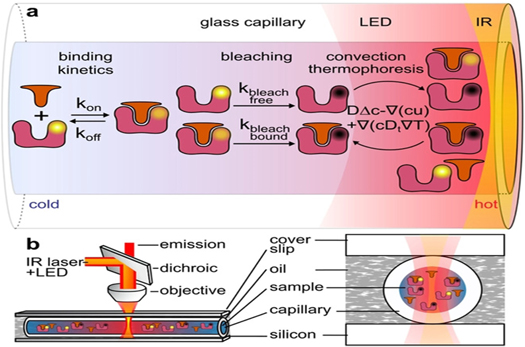
Usually 16 thin glass capillaries will be used for the measurement. The first 15 capillaries will be filled with solutions containing the same concentration of the intrinsic fluorescent or labeled fluorescent ligand but a decreasing concentration of non-fluorescent ligand molecules in a serial dilution from capillary 1 until capillary 15 (See figure 1). The last capillary (capillary number 16) will contain the same concentration of the target molecule as all the other 15 capillaries but without any ligand [47].

Figure 1. A representative diagram showing the process of measuring in microscale thermophoresis. IR laser beam focused on a capillary containing the target molecules induce temperature gradient. Ligand concentration is highest at tube 1, and this decreases until zero at tube 16. Unbound target molecules escape faster than bound molecules resulting in fluorescence drop.
Download figure:
Standard image High-resolution imageAs shown in figure 1, only one capillary tube is tested at a time. The detector moves then to the next capillary to record another plot and this is repeated ideally 16 times over the 16 capillaries in the tray. The reading starts with capillary No. 1 with the highest concentration of the ligand and ending with capillary 16 with a target molecule without ligand.
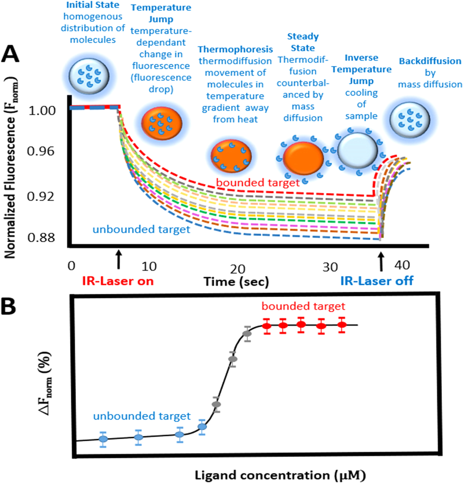
The MST movement profile includes four main phases. The movement profile of molecules in MST is schematically represented in figure 2(A) as normalized fluorescence percentage (F%) against time in second. Four main phases can be distinguished in the movement profile of MST. In phase 1 which last in about 5 seconds, the starting fluorescence will be identified before the start of the temperature gradient. This initial fluorescent before heating will be constant for all samples with and without ligand. Phase 2 is a fast phase that takes about 100 ms and represents the temperature jump phase. It indicates fluorescence change before thermophoretic movement (T-Jump temperature-dependent change of fluorescence). In this phase the fluorescence decreases initially due to heat-defendant change of fluorophore quantum yield. Phase 3 over several seconds represent the thermophoresis phase. During this phase the fluorescence decrease due to thermophoretic movement of the fluorescent target molecule away from heat in a temperature gradient until the steady-state is achieved. The steady-state indicates that the steady state thermo-diffusion is counterbalanced by mass diffusion. Phase 4 over about 5 s represent a reversed T Jump (inverse T-Jump or fluorescence recovery) IR switch off (or inactivation) result in back diffusion of the fluorescent molecules [6]. The cycle to complete the 4 phases would take about 20 seconds which is enough to reach the steady-state.

Figure 2. Representative diagram showing (A) the MST movement profile; phase 1 is initial homogenous state, phase 2 is induced temperature gradient through IR switch on, phase 3 represents thermophoretic movement, and phase 4 represent IR switch-off and reversed T jump; (B) the binding curve representing the ligand concentration versus the fluorescence drop. Modification from reference [6].
Download figure:
Standard image High-resolution imageLooking to the overlaid curves obtained by scanning the 16 capillaries the one at the top represent the capillary with the highest concentration of ligand (capillary No.1) and the one at the bottom represents the capillary without ligand and with only the target unbounded molecule (capillary No. 16) because unbounded molecule moves faster and thus the drop of fluorescence is faster as shown in figure 2(A).
Figure 2(B) shows the plot of normalized fluorescence against concentration of the ligand for the determination of the binding affinity between target and ligand. The kD value can then be determined from this curve. The smaller the kD value the greater the binding affinity of the ligand.
3. Label free versus labeled microscale thermophoresis
As mentioned before, MST is depending on the fluorescence of the target molecule to follow its movement. The fluorescence of the target molecule can best be an intrinsic fluorescence or a labeled fluorescence obtained via labeling with a fluorescence reagent. Different versions of MST instruments are available to best suit each case including label free instruments, labeled instrument, or instrument for both.
MST can in some cases work without labeling as other biophysical label free techniques. A label free target molecule represents an endogenously labeled molecule e.g. a protein with sufficient aromatic amino acid residues as tryptophan (intrinsic protein UV-fluorescence) or a small fluorescent molecule to conduct label-free thermophoresis. The use of label-free MST is unsuitable for serum or cell lysate which contain high background UV-florescence produced by the proteins in the sample [6].
A common disadvantage of label free thermophoresis is that protein-protein interaction cannot be measured because most proteins have enough aromatic residues to induce an interfering signal [48]. Labelled thermophoresis is the other alternative when the target molecule is nonfluorescent or is showing week fluorescent. An exogenously labelled target molecule with a covalently attached fluorophore in red or blue channel is then used to conduct the MST [31, 44, 49]. The labeling reagent my bind to other sites of the target molecule in what is referred to as (non-specific binding) might interfere with the binding of the ligand. In principle any of the two interacting partners could be labeled, but usually better to label the target large molecular weight protein. If the ligand is very small in size labeling it with a larger size dye and with a larger size anchor may change its properties [6]. Labeling is usually conducted using a fluorescent dye and a cross-linker to covalently bound the dye to a specific function group of target protein (e.g. N-terminal, primary amine of lysine side chain, arginine guanidino group in reduced cysteine residue, C-terminal. Unreacted excess dye should be removed before starting the experiment. Slide-A-lyzer-dialysis-cassettes or desalting column are two common ways to remove excess dye [50].
It is worth noting that, the exact position of labeling is not important for MST but important is just not to block or steric hinder the binding site of the ligand [6]. A site-specific and high-stoichiometric labeling should best be conducted. Heterogeneous nonspecific labeling would lead to a biased estimation of the binding affinity between target and ligand [49, 51, 52].
4. Sample loading and instrument running
Instruments with different detector types are in operation to conduct MST. Monolith NT.115 use visible light for fluorescence excitation with three LED-filter combinations, blue (excitation 460–480 nm, emission 515–530 nm), green (excitation 515–525 nm, emission 560–585 nm) and red (excitation 605–645 nm, emission 680–685 nm). Monolith NT.115Pico can detect lower concentrations in picomolar of red-emitting fluorophores. Monolith NT.LabelFree is used to detect the UV-fluorescence of proteins using 280 nm as excitation wavelength and 360 nm as emission wavelength. Monolithic NT.Automated uses two detectors for high throughput screening [6, 53].
To conduct the experiment an optimal fluorescence range and an optimal ligand concentration range should first be selected. Usually 16 capillaries (1 μm inner diameter) are loaded in the tray, each filled with constant concentration of the target fluorescent molecule and a decreasing concentration of the ligand (visible) non-fluorescent partner molecule the so-called serial dilution. First capillary holds the highest concentration of the ligand (where all binding sites of the target molecule are saturated with the ligand) and the one before the last (capillary no. 15) the lowest concentration of the ligand. The last capillary (capillary no. 16) is usually holds only the target molecule without ligand.
A standard glass capillary can be used for the measurements, however, adsorption of target or ligand to the capillary surface can be hindered by using capillary coated with polymers or by using buffer additives like detergents or proteins [6]. Practically about two thirds of the capillary is usually filled by dipping the end of the capillary in the tube containing the targeted solution. The capillary is then inverted to position the solution in its center and then placed in the capillary tray, which will be placed at the end in the instrument to perform the measures after loading the 16 capillaries. To avoid possible sample evaporation if experiments are to be performed over long time, the ends of the capillary can be plugged (sealed) e.g. with wax.
The excitation sources Light Emitting Diode (LED) power cab be adjusted based on fluorescence signal intensity. IR Laser power has wavelength of 1480 nm. The IR laser is focused in form of microscopic spot of about 50 μm in diameter on the sample in a volume of about 2 nl of the sample placed inside a glass capillary containing about 4 μl. IR laser induces the temperature increase thus temperature gradient △T of 2 °C–6 °C. Target molecules move away from elevated temperature and then diffuse back when the laser is switched off. Measures to the set are best repeated 3 times to take the mean and SD and ensure better reproducibility of measurements. The system software can provide the average and standard deviation.
5. Estimation of binding parameters
5.1. Steady-state binding affinity, KD
MST shows good capabilities to estimate the three basic binding parameters; binding affinity (pM to mM), binding kinetic and thermodynamics. Steady-state binding affinity KD indicates the strength of interaction and can directly be estimated through titrating different concentration (in molar ratio) of a non-fluorescent (optical visible) analyte (ligand) against a fixed concentration of either intrinsically fluorescent or labelled fluorescent target molecule. Analytical software is available for rapid data evaluation and graphical presentation of a binding curve and estimation of the binding affinity. Affinity binding KD values in millimolar to picomolar range have been reported noting that a smaller KD indicates higher binding strength between the two partner molecules (ligand and target) [8].
The plot of thermophoresis signal expressed as normalized fluorescence (Fnorm) against the concentration of the ligand (e.g. in micromolar or nanomolar) generates a binding curve which can give the binding constant. One should choose however between two fitting models available in the analysis software to fit the binding curve. Many bimolecular interactions use a simple model featuring the assumption of 1:1 binding interaction. For interactions involving multiple binding sites on target, the Hill equation model is used to calculate the concentration-dependent EC50 value. EC50 represents the effective dose of the ligand at which half of the target molecule are in bounded state [54]. The possible observation of biphasic curves for MST measurements with low and high KD values (<10–9 M and >10–8 M) might indicate the formation of different stoichiometry complexes [55].
Tso et al [54] proposed two additional MST analytical models assuming 1:2 binding scheme. The first assumes two microscopic binding constants KD1 and KD2. The second assumes symmetry in the bivalent molecule, culminating in a model with a single macroscopic dissociation constant (KD, M) and a single factor (α) that account for apparent cooperativity in the binding. Sometimes the conditions suitable to one biophysical method cannot be applied with another method so the use of at least two methods is recommended to have a better view about the binding affinity [56]. Recently, Sullivan et al [17] used surface plasmon resonance and biolayer interferometry in addition to MST to confirm a good binding affinity in nanomolar range for aptamers against β-Conglutin Allergen. In some cases, the obtained KD values from MST were in agreement with that obtained by other biophysical techniques [6]. MST is complementary used mostly with Surface Plasmon Resonance (SPR) and to less extend with Isothermal Titration Calorimetry (ITC) [57].
Sullivan et al [17] investigated the bimolecular interaction of aptamers against β-Conglutin Allergen using complementary techniques of MST, SPR and biolayer interferometry to confirm the nanomolar affinity. In one recent study to evaluate the binding of four bromobenzotriazoles to the catalytic subunit of human protein kinase CK2, MST gave a KD values in good agreement with that obtained by Isothermal titration calorimetry (ITC) [58].
In a recent study Vallejos-Sánchez et al [21] used MST, NMR and ITC to confirm no binding between pyrazinoic and ribosomal protein S1. Molecular dynamics and docking simulations are also used as complementary to identify the structural features that control affinities of binding [59, 60]. Usually good correlations are obtained between the computed KD value and the MST-derived kD values [61, 62]. High affinity and specificity are usually required for a certain ligand to be used for diagnostic or therapeutic application orthogonal competitive complementary techniques.
5.1.1. Variability in KD
The variability in binding KD may change in response to different biosensor type, surface chemistry and buffer composition may change binding behavior. These three techniques MST, SPR and ITC have different signal read-out or change, while SPR depends on change in refractive index, ITC depends on change in temperature (heat change) and MST depends on change in fluorescence intensity [63]. Compared to buffered salt solution that lack many components of original fluid to mimic in vivo conditions, measurements of KD using cell extract result usually in higher values compared to buffered salts. That is to say that lower binding affinity is observed for real sample (original fluid). Christoph et al [9] attributed this to possible competitive interaction from other components in the original fluid or different viscosity, pH or ionic strength. One study showed that the MST-derived KD value is lower than that of SPR-derived KD value which was attributed to methodological difference having labeling in MST and immobilization in SPR [27].
Sass et al [27] showed that for aptamer –DNA the dissociation constants determined with MST were always lower than that from SPR, and especially for the truncated aptamer they differed by two orders of magnitude. They suggested that the methodological differences (MST labeling) and SPR immobilization may create differences in dissociation constant.
5.2. Binding kinetics
Binding kinetics is particularly important to determine when investigating biomolecular interactions for therapeutic purposes. Binding kinetic is represented by estimating the values of association rate constant ka (also known as kon ) and dissociation rated constants KD (also known as koff). kon indicates the speed of association between the ligand and target and koff indicates the duration of stability of the complex. A good wished potential therapeutic candidate would have fast kin and slow koff. Proper values are also considered for diagnostic purposes based on the aimed application. Determination of the kinetic parameters typically requires analysis using multiple concentrations.
MST has not been recommended to allow obtaining kinetic information and measurement of kon and koff rates [64, 65]. However, other authors showed the ability of MST to estimate the binding kinetic. Thus indicating that MST is not only particularly successful in characterizing interaction in steady-state equilibrium but it can also analyze dynamic binding kinetics. In one study, MST has been used to obtain kinetic binding data of the Argonaute-2 protein with a miRNA, which showed a possible RNA-induced silencing complex (RISC)-mediated turnover of inhibited miRNAs [66]. In another study, Jerabek-Willemsen et al [67] demonstrated that MST can quantify enzyme kinetic.
Julian et al [68] have recently introduced the term kinetic microscale thermophoresis (KMST). Their research proved that MST is a cost and time effective technique that can be extended to measure reaction kinetics besides KD in one experiment. The researchers used the temperature jump technique, making slight modification in the conventional MST hardware. As shown in figure 3, The slight modification in the MST hardware was set in order to effect enough increase in the sample's thermal dissipation for deduction of kinetic relaxation from the fluorescence intensity. Julian et al studied the DNA hybridization and its kinetic parameters. KMST proved superiority over immobilization techniques, such as SPR and BLI, for determination of reaction rates both in time and cost. Moreover, KMST overcomes the alteration in physical or chemical characters of biomolecules due to immobilization.
Figure 3. KMST hardware modifications. Capillaries are contained in silicon wafer and immersed in oil. Reproduced based on the terms of the creative common attribution license from reference [68].
Download figure:
Standard image High-resolution image
5.3. Thermodynamics
Thermodynamics help to understand the principles of interaction. Negative value of Gibbs free energy (△G) derived by enthalpy or entropy would indicate spontaneous occurrence of the interaction between the ligand and the target. The enthalpy parameter (△H) shows the change in energy due to noncovalent interaction or hydrogen bond or van der Waals interaction between target, ligand and the solvent. On the other hand, the Entropic counterpart (△S) shows the degree of freedom of a system as a global thermodynamic property.
MST can estimate the thermodynamic of interaction by variation of assay temperature [67]. In a study investigating the interaction between Fe(III) and anthocyanin, MST estimated ΔG < 0, ΔS < 0 and ΔH < 0 thus indicated that the chelation interaction is exothermic and spontaneous and involve van der Walls and/or hydrogen bonding [5].
6. Contribution to the study of biomolecular interaction
In 2018, El Deeb and his group mentioned in a review the versatile application of MST for various kinds of biomolecular interaction [8]. MST has been used for the characterization of binding events either as a sole technique [69] or in addition to other orthogonal techniques to evaluate binary and multiple biomolecular interaction [4]. In some works, MST has been used to confirm molecular interactions revealed by molecular docking [70] in other studies molecular modeling and docking studies are used to support MST results [25, 71, 72]. The technique found good applicability in high throughput analysis to screen large libraries within a reasonable fast time with high precision [22, 73].
MST can be used to study protein-protein biomolecular interactions (PPBI). In 2019, Lee et al [74] used MST to study the affinity of phosphorylated BECN1 protein to the anti-apoptotic proteins; BCL2 and BCL2L1. Their study proposed that phosphorylation of BECN1 at threonine (T108) within its BH3 domain could enhance electrostatic interactions with the histidine residue on BCL2 and BCL2L1. MST was used in determining KD between phosphorylated and non-phosphorylated BECN1 proteins with BCL2L1. The lowered KD values proved the higher affinity to the phosphorylated BECN1 which indicated its ability to regulate autophagy. Lee at al also studied the effect of local environment on binding strengths. MST proved that presence of surfactants (Triton X-100) caused about 6 folds' increase in the affinity between phosphorylated BECN1 and BCL2L1 proteins.
Berleth et al [75] studied the perception and transmission of signals in plants mediated by PPBI. To quantify this interaction in the plant ethylene pathway, MST was applied. The authors suggested that ethylene receptor subfamily-II, ETR2, has a role in PPBI-mediated signal transfer. The proteins of downstream ethylene pathway, CTR1 and EIN2, were the ligands. The reported research also evaluated the stability of type-II receptor complexes as compared to type-I receptors isoforms. MST calculation results of dissociation constants proved higher stability of the ethylene type-II receptor complexes indicated the importance of these subtype receptors in signal transduction. The authors used purified recombinant ETR2 and EIN2 proteins, which were labeled first with Alexa Fluor 488 succinimidyl ester before MST experiments. Plant in vitro studies using the biophysical technique, MST, enabled with ease the quantification of KD and hence the binding affinities.
In 2020, Magnez et al [76] proposed that MST is a powerful and more reliable tool for measuring PPBI as compared to surface plasmon resonance and isothermal titration calorimetry. The authors studied the binding interaction between PD-1 and PD-L1 to calculate the values of their dissociation constant KD, which suffered controversy when was measured by the mentioned techniques. Moreover, the KD results were calculated for human PD-1/PD-L1, murine PD-1/PD-L1, and cross-species murine PD-1/ human PD-L1 in such a way that proved similarity regardless of such variations. The application of their proposed MST protocol enabled for the first time, as claimed, the determination of PD-1/PD-L1 affinities during tumor escape. This protocol also using MST could accelerate the characterization of PPBI for studying newly developed molecules effects on modulating the affinity of PD-1/PD-L1 binding as new strategies that target immune escape for oncology.
Unlike normal cells, cancer cells depend on glutamine for their metabolic activity. So, the enzyme aspartate transaminase 1 (GOT1) is important to catalyze the anabolic activity of cancer cells in the generation of nicotinamide adenine dinucleotide phosphate (NADPH). In 2019, Sun et al [77] claimed the activity of a natural compound isolated from a culture of Aspergillus terreus, Aspulvinone O (AO), in blocking GOT1 protein. Pancreatic ductal adenocarcinoma (PDAC) cells were used throughout the experiments since they rely on GOT1 in rewiring glutamine metabolism. Virtual docking of AO revealed that it can bind effectively at GOT1 active sites by several hydrophobic interactions. MST was used by the authors to calculate the affinity between AO and GOT1 in order to verify their findings. Recombinant GOT1 was labeled and mixed with AO in serial dilutions to calculate their KD. The KD results proved the specific binding of such substrate to homo and murine GOT1 indicating the inhibitory effect and suppressing PDAC cell proliferation.
The technical challenges facing the study of protein-protein interactions in membrane proteins are mainly bases on the small amounts of membrane proteins available and the need for detergents use. That's why these studies were based mainly on peptides corresponding to the full-length proteins. MST has the capability of facing those challenges. A recent study compared protein-protein interactions for human aquaporins (AQP), which are responsible for gating and trafficking water transport along the cellular osmotic pressure [38]. The research studied the binding of LIP5 and CaM ligands to both; full length AQP2 and AQP0 as well as peptides corresponding to the AQPs binding sites, respectively, using MST. MST results proved that although such peptides can locate the binding sites and the binding interactions, the study of full-length proteins doesn't the important parameters of allosteric regulation and the cooperativity between binding sites. MST estimation of KD values for full length AQP0-CaM and AQP2-LIP5 versus their peptides-ligand interactions were found differing and the results were comparable to those obtained by fluorescence anisotropy technique. This study highlighted the importance of studying full length proteins instead of their corresponding peptides to demonstrate other contributions such as binding cooperativity as shown in AQP0-CaM results.
The involvement of MST in the characterization of designed peptide mimetics were reported in some research articles. In 2019, Sencanski et al [28] used MST to characterize β2-adrinergic receptor (β2-AR) related nanobody derived peptide (NDP). The selected NDP of Nb71, designated as P3, interaction with agonist activated and unstimulated β2-AR using MST. Estimated EC50 results revealed that P3 had 10 folds higher affinity to agonist activated β2-AR activated by isoproterenol. In the same research, P3 ability to interfere with the agonist activated β2-AR with β-arrestin 2 was studied by MST. The results showed low evidence of P3 interfering capability. Dose-response curves derived by MST for P3 biomolecular interaction with β2-AR were an easy and reliable tool for prediction and verification of the newly designed ligands.
Also, MST measurements were utilized to study antigen binding to colloidal saponin associations as new potential adjuvants for vaccines. In the reported study, Carolin et al [78] studied the binding of labeled ovalbumin model antigen and saponin (SBb, SAb, ß-escin)—cholesterol ligand binary system. After application of the temperature gradient, higher relative fluorescence than ovalbumin alone was observed. The fluorescence increase was due to the labeled ovalbumin altered thermophoresis behavior after binding with the ligand. These results proved the binding between ovalbumin, model antigen, and saponin– cholesterol ligand binary system. As a result, both soyasaponin in its pure form or in formulation were proven to be promising adjuvant systems for the intradermal vaccine application
Furthermore, MST was utilized to assess the binding interaction between flavonoids and proteins. In 2020, MST was also involved to quantify the dissociation between bovine lactoferrin ligand (BLF), labeled with RED-NHS dye, and one of the two flavonoids dihydromyricetin (DMY) and myricetin (MY) and binding impact on the in vitro radical scavenging activities of the complexes [30]. KD for BLF-DMY and BLF-MY interactions of were found to be 109.8 ± 4.96 μM and 17.65 ± 0.34 μM, respectively. MY had smaller KD value reflecting its higher affinity for BLF. Thus, BLF was found to be 6.22 times stronger transporting capacity than DMY leading to greater absorption and bioavailability of MY.
One of the effective therapeutic treatment methods in Alzheimer's disease (AD) is the interference in β- amyloid protein (Aβ42) aggregation and its related cytotoxicity. Limeng et al [79] found that chitosan oligosaccharides (COS) had excellent blood-brain barrier (BBB) penetration capability and could interfere the β- amyloid protein (Aβ42) aggregation resulting in reduction of its related cytotoxicity. MST was applied to study the binding affinities of Aβ42 with COS COS monomers. Cyanine 5 (Cy5)-labeled Aβ42 adsorption of onto the capillary walls of MST was studied prior to experiments indicating no self-aggregation or absorption on the wall. In the following experiments, different concentrations of the Cy5 labeled Aβ42 and non-fluorescent labeled COS mixture as binding ligand were used. The fluorescently labeled molecule thermophoretic movement changed after binding to a non-fluorescent ligand. The relative fluorescence intensities between the bound and unbound state were recorded. The results revealed that a single binding case between Aβ42 and COS mixture with KD of (3.76 ± 0.34 μM) was observed. Also, MST was used to study the binding affinity between COS monomers with oligosaccharides DPs [3–6] and calculating the binding affinities (KD). The binding affinities were found to be DP-dependent. COS monomer binding affinity with DP6 was found to be the strongest due to having the lowest KD (2.09 ± 0.87 μM) and highest Ka (5.95 ± 2.48 × 10⁵ M-1). This research highlighted COS potential role as novel therapeutic agents for AD treatment.
The study of the binding of αS1-casein, human milk protein, is very important to understand its role in lifelong immune reaction towards αS1-casein. Thorsten et al [80] used MST to characterize and quantify the binding of the milk protein αS1-casein as a binder of the TLR4 ecto domain and dissociation constants (KD) were calculated. It was found that αS1-casein could bind with TLR4 alone (2.7 μM KD), with the cofactors of TLR4 MD2 (0.3 μM KD) and CD14 (2.7 μM KD). Also, LPS-TLR4/MD2 binding constants were estimated (8.7 μM). It was found that higher concentrations of TAK-242 (10-times) needed to inhibit αS1-casein nad LPS- induced effects. Furthermore, αS1-casein was found to be stronger ligand of TLR4 than LPS. αS1-casein induced effects were found to be CD14-dependent unlike LPS.
MST was also used to study the interactions between insecticides and some chemosensory proteins that caused insect survival through insecticide exposure resulting in insecticide resistance. Xiao-Qiang et al [81] studied the binding interaction between Diaphorina citri odorant binding proteins (DcitOBP2) and different insecticides. The DcitOBP2 was first bound to fluorescence dye (KD = 16.06 nM) then serial dilutions were made in three different neonicotinoid insecticides. The DcitOBP2- neonicotinoid insecticide binding behavior was dose-dependent. DcitOBP2 binding affinities KD were 62.39 nM with imidacloprid, 128.68 nM with thiamethoxam and 186.98 nM with dinotefuran indicating strong binding behavior. On the contrary, no binding affinity was observed for DcitOBP2 with non-neonicotinoid insecticides (fenpropathrin, abamectin and chlorpyrifos). The purified recombinant protein of DcitOBP2 expressed in Escherichia coli showed strong in vitro binding activity (KD = 62.39 nM) to imidacloprid using MST. Imidacloprid is a systemic insecticide that acts as an insect neurotoxin and belongs to a class of chemicals called the neonicotinoids which act on the central nervous system of insects.
The interaction of PIRT (the phosphoinositide regulator of TRP) with TRPM8 (the cold and pain human sensor) and with the signaling phosphoinositide lipid PIP2 were studied by Nicholas et al using MST [82]. MST confirmed that hPIRT interacts with both PIP2 and the hTRPM8-S1S4 domain and the hPIRT affinity to interact with the hTRPM8-S1S4 domain was 35-fold higher (lower KD) than with PIP2. Also, the interaction between hTRPM8-S1S4 and hPIRT decreased 7-fold in presence of saturating PIP2 (higher KD) values. This paper succeeded to suggest the mechanism by which TRPM8, PIP2, and PIRT could form a regulatory complex and the PIRT modulation of TRPM8 was attributed to the accessible concentrations of PIP2reaching TRPM8.
MST was utilized to confirm the strong binding affinity of metallacarborane to crude snake venom proteins (KD in nM range) but, moderate binding affinity between metallacarborane and the short single-stranded DNA (KD in μM range) [83].
Kamal et al [84] used MST to evaluate the binding affinity of HIV-1 capsid (CA) inhibitors to the cyclic peptide (Pep-1) and capsid inhibitor (PF74). The movement of molecules was detected using a covalently attached fluorophore. It was found that the binding affinity of CA with Pep-1 (KD = 32 ± 3 nM) was 7-fold higher than PF74 (KD = PF74 212 ± 7 nM).
MST was applied as a tool for prediction of the binding affinities between melanin with drugs as this binding had a great effect on medications efficacy and toxicity especially in diseases of ophthalmic and pigmented tissues disorders (e.g. melanoma) [46]. MST used the melanin natural auto-fluorescence to determine dissociation constants (KD) of melanin nanoparticles with eleven drugs. The results revealed that the cited drugs could be classified into four categories according to the strength of binding (KD values). The four categories were extreme (penicillin G, chloroquine), high (levofloxacin, terazosin, papaverine), intermediate (propranolol timolol, quinidine, nadolol,), and low binders (methotrexate, diclofenac, atropine).
Guangcheng et al [85] used MST to evaluate anti- Tomato chlorosis virus (ToCV) activities of some novel dithioacetal containing agents through the study of their interaction with ToCV coat protein (ToCV-CP) as a novel target. The results indicated that there was a strong binding of the agents C5 and C22 to ToCV-CP. Their binding constants were 0.24 and 0.25 μM, respectively.
MST was used to assess mRNA cap interaction with various proteins involved in some therapeutically important mRNA-related processes using designed fluorescent probes [86]. Michal et al studied the interaction between newly synthesized m7Gp3G cap derivatives with two cap-binding proteins, decapping scavenger (DcpS) and eukaryotic translation initiation factor (eIF4E) using different fluorescein-labeled probes. All probes KD values were found to be in nM range. Probe 12 could tightly bind DcpS (14 nM, KD).
Gao et al [87] determined the binding of RNA (SH7) to human respiratory syncytial virus (HRSV) M2–1 protein in all-atom simulations using MST. The results revealed that the RNA interactions with two M2–1 domains which were the core domain (CD) and zinc-binding domain (ZBD) were independent. This research provided a structural basis for recognition of RNA using HRSV M2–1.
Macut et al [88] used MST to screen a library of small peptides as ligands against a target enzyme regulating intracellular fructose-2,6-bisphosphate level.
The effect of osmolytes and some anions on the interactions between supported lipid bilayers (SLBs) and NPs was investigated using MST [89]. 1,2-dioleoyl-sn-glycero-3-phosphocholine (DOPC) as lipid component and 60 nm-carboxylated polystyrene as NPs were chosen to develop the cited model. MST was applied for binding characterization between DOPC vesicles or NP and osmolytes. The results revealed that osmolytes could protect SLBs against the induced disruption caused by NPs. The combination of kosmotropes (F_ and TMAO) had higher efficiency than chaotropes (NO_3 and urea) in weakening NPs and SLBs interaction.
Nikola et al [90] has studied the sense—antisense peptide interactions using MST under different binding circumstances. These interactions could affect peptide ligand—receptor systems selection.
Bharambe and Ramachandran in their book chapter [91], applied MST applied to determine human DSPs-lipid interactions for target cardiolipin in liposomes and estimating KD. 0.1 μM of full-length Drp1 binding with 100% CL liposomes had 36.3 ± 14.4 μM KD. While, higher concentration (0.9 μM) had 12.7 ± 4.5 μM KD. No interaction was observed for Drp1 VD with control 100% DOPC liposomes.
MST shares the importance among group of analytical approaches aimed at characterization of protein-peptide binding. In 2019, MST was used together with fluorescence, NMR, Surface Plasmon Resonance (SPR) and MALDI-TOF/UPLC-HRMS to test a hypothesis about the effects of amyloid β peptide (Aβ) in clearance pathways of Ubiquitin (Ub) proteins in Alzehimer's disease patients. MST served to calculate KD values of Aβ-Ub competitive binding which in turn suggested the peptide role in the pathological failure for clearance of proteins accumulated in brains of Alzehimer's disease patients [92].
MST helped in the evaluation of newly developed small clinically important peptides, aptamers. Aptamers are oligonucleotide or peptide molecules that can bind specifically to target molecules which have evolved in their importance in basic research and clinical purposes. The main limitation that faced the development of aptamers was lacking suitable and sensitive methods for their evaluation. MST was evaluated by Svobodová et al [93] as a rapid tool with high throughput in analysis of binding affinities and kinetics between aptamers and target small molecules. Their research studied the binding specificity and affinity of several 17β-estradiol aptamers. KD constant were calculated using studied aptamer 17β-estradiol, testosterone, progesterone and androstenedione. MST results were compared to those obtained by two other approaches, Surface Plasmon Resonance (SPR) and Apta-PCR affinity assay (APAA). The agreement between obtained results proved the suitability of MST as fast and reliable approach in evaluation of the clinically and diagnostically important aptamers.
MST can be used to confirm RNA-ligand binding. Wang et al [94] conducted a research study on accumulation of antibiotic resistance genes for class 1 integrons. Class 1 integrons play an important role in antibiotic resistance dissemination since they have the ability to capture the resistance gene cassettes and insert them into specific sites to be transcribed. MST was used to measure aminoglycosides' binding affinities to riboswitch RNA. Several aminoglycosides (including tobramycin, amikacin, gentamycin, ribostamycin, and neamine) were used as ligands to test their binding affinities as represented by KD values. Aminoglycosides that had higher affinities to riboswitch RNA were able to induce the expression of reporter genes, while those that had low affinities couldn't. MST was used to provide in-vitro biomedical evidence for such hypothesis and its results were augmented by covariance analysis.
In 2018, a hypothesis was tested to model pontocerebellar hypoplasia type-1B (PCH1B) neurological disorder. The researchers tried to proof a link between mutations in EXOSC3, a putative RNA-binding structural cap protein, and PCH1B. EXOSC3 is one of three cap protein which helps guiding RNA into the RNA exosome. MST helped to test the hypothesis at different levels throughout the research [95]. First, MST was used to test the affinity of EXOSC3 to the long and short G-rich RNA sequences. Results showed higher KD values between EXOSC and long than short G-rich RNAs. MST was then used to screen the binding of about 50000 small molecules to EXOSC3 to assess their abilities to inhibit RNA binding by EXOSC3. Those experimentations that involved MST lead to the discovery of a new compound that disrupted EXOSC-RNA binding, which was called (ERD03). ERD03 was proved to bind specifically to EXOSC3 causing the disruption of binding between EXOSC3 and RNA in a manner dependent on its concentration. In such a way, ERD03 induced PCH1B-loke phenotype causing zebra fish embryos to develop abnormal curved spine. Mutations in EXOSC3 impaired its RNA-binding affinity as proven by MST results tested on both long and short G-rich RNAs.
Phospholipids interactions with proteins are important to be investigated since they comprise triggering of several intracellular events. MST was used to study such effects as suggested by Tindall et al [96] when studied the binding of vaspin, a protease inhibitor, to membrane phospholipids and polyphosphates. KD results to labeled vaspin, as calculated using MST, revealed the high affinity of binding to polyphosphate 45 resulting in activation of vaspin in a heparin-like manner. These findings elaborated the importance of vaspin-phospholipid and polyphosphate binding in membrane trafficking parameters affected by obesity such as glucose tolerance and inflammation of adipose tissue.
The difference between in-vitro and in-vivo protein interactions with developed drugs were proven using MST. This difference is crucial to provide reliable conclusions about newly developed drugs [9]. This fact was explored by Wienken et al by determining the affinity of quercetin, an inhibitor small molecule, to its protein kinase PKA in buffer and human serum. The 400 folds reduction in affinity of quercetin-kinase in human serum than in buffers indicates the important effect of biological matrices in drug developments. The study highlighted the significant competitive binding effects of several components originally present in the biological matrices that are lacked in the artificial in-vitro buffers which may lead to wrong decisions about candidate drugs.
MST isn't only used to study affinities, but also used to study cellular DNA functions and folding processes. Zhang et al used MST to study the DNA folding and unfolding pathways stabilized by cations [97]. MST helped in monitoring the changes in charge and size of Guanine quadruplexes (G4s) when bound to K+ cation. Thermodynamic parameters had been able to be investigated and used to identify two predicted G4 folding pathways that were energetically favored.
MST was used to test the binding affinities between small molecules and proteins in several publications. MST was recently used to detect the interactions of variety of small molecules to group of fibrillar protein aggregates. Fibrillar protein aggregates are identified in the human brains of neurodegenerative diseases such as Parkinsonism and Alzehimer's disease [98]. α-syn and tau fibrils were used as target proteins for several small molecules to calculate the KD and Ki using MST. The aforementioned study made use of advantages of MST as highly sensitive in presence of small amounts of samples, however, providing consistent results.
During COVID-19 pandemic, MST had beneficial use to investigate and/or affirm possible regimens. In 2022, Papaj et al [99] used MST to investigate the in silico studies that suggested three factor Xa inhibitors, namely Apixaban, Rivaroxaban and Betrixaban, as possible potential inhibitors for SARS-CoV-2 protease (Mpro). Experimental results offered by MST indicated that the drugs under study had no therapeutic effect on COVID-19. No binding affinity was found between Rivaroxaban and the target enzyme. Apixaban, and Betrixaban could be bound to the target only at high concentrations which is not practical due to their low solubility. Such high concentrations cannot be achieved in plasma; therefore MST results excluded the suggestions of in silico studies.
The development in MST applications is dynamic and growing fast. Optimistic research article was recently reported for the use of MST in the study of acid-base equilibrium and determination of pKa values [100]. Nowak and Wo´zniakiewicz reported direct relationship between deprotonation of Fluorescein isothiocyanate and thermophoresis results. Acid-base equilibrium investigated on the analyte under study using MST showed markable thermophoretic response caused by the deprotonation step. Their pKa findings were consistent with reported results. Measurement of pKa values for drugs using MST magnifies the analytical potential of this technique. pKa values of drugs are important in their pharmacodynamics and pharmacokinetic studies. The simple operation, sensitivity, lower sample utilization, beside its ability to be carried out in biological compositions, increases the importance of MST as analytical tool in this area.
The investigation of the mechanism by which the cardiac isoform of myosin-binding protein C (cMyBP-C) is myosin-based regulated was performed using MST [101]. A new model was established by extensive mapping of cMyBP-C interactions by MST using fragments of β-cardiac myosin.
A recent research article investigated the binding activities of some small molecules to a protein receptor in order to identify and correlate their pharmacologic effects. Cereblon (CRBN) is a substrate protein receptor that was identified as target for immunomodulatory drugs (IMDs) derived from thalidomide molecule. MST was used to measure the binding affinities for a group of thalidomide derived compounds, fluorinated analogues, and some related metabolites to CRBN and correlate their antiangiogenic effects. The results of Ki and IC50 obtained excluded the correlation between CRBN binding affinities and the inhibitory effect to angiogenesis, and hence thalidomide analogues antiangiogenic effects remained to be identified [102].
It is worth noting that, small molecular weight ligands often did not change the charge or size of a large molecular weight target protein upon binding but can induce weak conformational change and change in hydration entropy due to the release of some water molecules on the protein surface thus changing the hydration shell. The change in such a thermophoretic parameter is enough to induce a change in thermophoretic signal to enable the estimation of binding parameters using MST.
Approximately 30% of all proteins in biological systems are metalloproteins and thus exploring the interaction between protein and metal ions is very important for to understand biological mechanisms, diseases and to design drugs [103].
Deferiprone as a target molecule shows good selectivity toward metal ions of about similar size and charge by binding some and showing no binding to others and giving different binding constant values for each metal ion [8].
7. Selected diagnostic and therapeutic applications involving MST aided characterization or MST based technology
MST has certain potential for therapeutic and diagnostic application. One important field is the development of biosensor. Biomolecules that are highly expressed and structurally stable at early stage of a disease are target for diagnostic purposes through the assignment of a proper ligand molecule that can bind to it in high affinity and selectivity. In vitro evaluation techniques to develop biosensors are used to characterize biomolecular interaction and identify the dynamic of the target molecule and define a good ligand that can be implemented in a proper biosensor technology. One best of a set of affinity ligands against candidate target molecule e.g. biomarker or antibody or antigen that can develop into a rapid test is usually selected. In addition to the key factors of high affinity and selectivity, sensitivity is also considered according to the intended application and the sample concentration. Low LOD are sometimes mandatory and can be a key player in different competitive assays [104]. For the development of biosensor system low KD ligands (high affinity to bind) that can also exert good selectivity are wishes sensor molecules.
Rapid diagnostic tests based on different technologies and either antibody-mediated or biomarker-mediated are based on bimolecular interaction characterization facilitated by techniques as MST and using sensor molecules as aptamers. Rapid and sensitive detection biosensor technologies are highly wished to replay other expensive detection platforms based on chromatographic separations or antibody-antigen assays [93]. Aptamer-based, antibody-based are powerful tools for fast biosensor devices.
It is worth noting that aptamer which are synthetic single-stranded DNA or RNA sequences are good ligand molecules which can be synthesized against certain target molecules generated usually using systematic evolution of ligands by exponential enrichment (SELEX) methodology to exert affinity and selectivity. Their ability to fold into tertiary structure offers them high affinity and selectivity against the target. They can usually achieve low KD values e.g. in micromolar range indicating their high affinity binding [105].
In case of insufficient affinity or selectivity a post-SELEX optimization approach to engineer high affinity variants is usually conducted [106]. That is usually followed by in-silico high throughput computational method for the screening of several generated aptamers to test affinity. The theoretical results are then followed by practical characterization where MST can play a powerful role to reveal agreement of the theoretical expected affinity [95, 107].
Due to the high charge of aptamers they can induce a strong thermophoretic response upon binding a target [9]. MST is currently a key player in studying biomolecular interactions involving aptamers as a sole technique, or together with other one or two orthogonal techniques, and/or with the support of molecular docking to compare a theoretical derived affinity binding value. Recently, MST has been applied for the development of aptamer-based recogongnision biosensor the so called MST-aptasensor [108]. In 2017 Skouridou et al [109] linked the binding data for a progesterone aptamer obtained from MST to the structural differences on the ring structure of a range of steroid in an approach referred to as aptatope mapping.
Moreover, MST was used to improve the clinically diagnostic tools. In a recent work [110], MST has been used to characterize the binding between Gd(III)-loaded fibrinogen aptamer (FA) chelate conjugate and fibrin. MST proved the high affinity between fibrin and the studied aptamer and their KD was calculated. Proving the high selectivity and affinity between fibrin and Gd(III)-loaded-FA using MST enabled its use for the magnetic resonance imaging targeted for identifying the location of blood clots where fibrin is a major constituent.
For the diagnosis of infections caused by Aspergillus and Penicillium species, McKeague et al [111] studied the binding affinity of 3 bio-recognition agents. Aptamers were reported for the detection of ochratoxin A, which is a mycotoxin produced as secondary metabolite from the mentioned species. MST findings recommended one of the three families of aptamers (Aptamer A08min) to be the ideal one for developing biosensors for the bio-recognition of ochratoxin A.
Being of valuable potential applications, aptamers which can be used for diagnostic, analytical or even therapeutic purposes should be studied. In an interesting study, MST was used to investigate the interaction pattern of aptamers targeting HIV-1 integrase enzyme [112]. Analogues to d(GGGT)4 aptamer, which had a basic site and replacing thymidines from the original sequence, were evaluated for their binding affinities to the labeled target enzyme. MST results concluded that MST is capable for assisting in the studies aimed for molecular modeling and docking.
MST is also helpful as analytic tool. It was used in optimizing specific and selective aptamers for the determination of Biphenol A which is one of the environmental endocrine disruptors [113]. The developed method had a low detection limit for the target biphenol A, besides being fast and simple. The developed method could be applicable for the analysis of water samples.
New diagnostic and prognostic data was evaluated for melanoma with the help of MST [114]. Sobiepanek et al proposed a new tool distinguishing melanoma progression phases based on lectin-glycan interaction.
8. Advantages of MST
MST technique has many advantages in estimating the binding affinities when compared with other related techniques. First, MST is simple, robust and fast technique which allows the quantitative and straightforward study of any type of molecular binding. MST possesses the advantage of high sensitivity as it depends on fluorescence measurement. Therefore, it allows the determination of very low concentrations even in nanomolar scale. Also, MST permits high throughput screening using very small sample size (microliter scale) [22], unlike the other biophysical techniques which require larger sizes such as ITC and SPR. The low sample consumption of MST (about 192 μl) is attributed to holding the samples up in sixteen 4 μl -capillary glass tubes in triplicates (16 × 4 × 3) as maximum sample size. In comparison with SPE, MST is advantageous in being simpler and being in-solution technique. SPR needs extensive optimization steps and target molecule immobilization, while MST needs simpler optimization, no immobilization steps resulting in performing the experiments in the solution itself. Hence, MST is a simpler immobilization-free technique which is considered as an alternative technique to SPR [115].
Moreover, the presence of the target biomolecule in a complex unpurified liquid (e.g. cell lysate, serum and plasma) does not represent an obstacle to use MST as it can be applied without the need to purify proteins from the matrix [49]. This advantage enables researchers to form a very close image to the in vivo interactions providing them efficient and precise interaction information. So, MST is considered as in vivo imitative technique in the fields of drug development and discovery, disease diagnosis and biomarkers interactions [6, 9]. Also, MST experiments do not involve any contact with samples so the risk of contamination is non-existent. In MST experiments, no restrictions are involved in selection of neither buffer nor partner molecular weight as MST can be performed using different buffers and no limits for molecules weight were found [48, 64]. MST instrument has an integrated quality control characterization part. Furthermore, binding study in MST does not depend on ligand size. The reason is the observable change in hydration shell or charge which occurs even in presence of small ligand size despite the unobservable size change. MST succeeds to work in presence of micelles and detergents which hinders the aggregation of proteins. Moreover, MST has the advantage of low sample consumption, which agrees to green analytical chemistry concepts [116, 117].
Nevertheless, it does not need any maintenance leading to decrease the cost of analysis. That means, MST is cost effective if compared with other expensive techniques such as isothermal titration calorimetry.
Last but not least, MST has multiple applications. It can be applied for the quantitative determination of target ligand interaction, studying the modes of binding such as dimerization competitions and cooperativity, getting kinetic binding data and EC50 assessment [66]. Also, it can be used for rapid antibody selection owing to performing the measurement using the cell suspension supernatant. It can be applied in the detection of protein unfolding [118]. Also, it is used to detect the binding mode occurring due to protein solvation entropy change accompanied with size and charge unchanged [119].
On the other hand, there are some major disadvantages for MST. The measurement of protein-protein interactions using MST cannot be allowed unless one of the interacting proteins does not possess much aromatic residues to give measurable signal [48]. MST is highly sensitive to any change in the molecular properties, such as size, charge, or conformation [67]. While Surface plasmon resonance (SPR) and bio-layer interferometry (BLI) techniques used to investigate biomolecular interactions are label-free [115], MST may require fluorescent labeling. If fluorescent labeling is required for the target, MST would represent a time consuming process [120]. The labeling itself may cause non-specific binding since MST cannot discern second binding site.
MST may require additional NMR or x-ray crystallography studies since it doesn't offer clear information about the binding location or stoichiometry [120]. Until now, there are only 2 models used for analyzing the data obtained by MST, 1:1 interaction model and Hill model, with the assumption that the ligand and labeled receptor has 1:1 molar interaction ratio. However, if the interaction have 1:2 molar interaction stoichiometry, these models would not serve in data analysis [54]. MST also has the disadvantage of a short working temperature range (20 °C–45 °C).
9. Future of MST
More future applications in the field of MST should be revealed progressively. MST is a simple and green analytical technique for monitoring the molecular interactions, therefore it's expected to have wider application in the quality control of biopharmaceuticals. The capabilities based in MST to identify any tiny change in conformation and structure of biomolecules, for instance upon thermal heating, enhance its role in enriching this scientific platform, such as thermal stability studies. Moreover, MST's role in measurement of the enzymatic activities would benefit more in the identification of formed products and assays. In a disclosing statement, much more insights in the capabilities of MST shall be revealed in the scientific work in the near future.
Data availability statement
No new data were created or analysed in this study.
Supplementary materials
The research article does not have electronic supporting information.
Informed consent statement
Not applicable.
Declaration
There is no conflict of interests to declare.
Funds
There are no funds to declare.